Nuclear Medicine
Nuclear medicine is the use of very small amounts of radioactive material to diagnose and, sometimes, treat disease. Nuclear medicine can provide accurate images of specific areas of the body; valuable information about how your body is working; and therapy to fight some diseases. Nuclear medicine can detect a wide variety of conditions and illnesses, such as arthritis, heart disease, cancer and infection
Patients Can Expect
- Minimal wait time
- Acceptance of most medical insurance plans
What to Bring
- Physician Order
- Current medical insurance card
- Drivers license or other government issued identification
- Wear comfortable clothing. (Avoid metal straps, buttons, zippers).
- You may be asked to change into a gown.
Click here for imaging locations.
Echocardiagram
(Echocardiography, Echo, Cardiac Ultrasound, Cardiac Ultrasonography, Cardiac Doppler, Transthoracic Echocardiogram, TTE)

Procedure Overview
What is an echocardiogram?
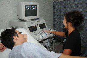
An echocardiogram is a noninvasive procedure used to assess the heart's function and structures. During the procedure, a transducer (like a microphone) sends out ultrasonic sound waves at a frequency too high to be heard. When the transducer is placed on the chest at certain locations and angles, the ultrasonic sound waves move through the skin and other body tissues to the heart tissues, where the waves echo off of the heart structures. The transducer picks up the reflected waves and sends them to a computer. The computer interprets the echoes into an image of the heart walls and valves.
Other related procedures that may be used to assess the heart include resting or exercise electrocardiogram (ECG or EKG), Holter monitor, signal-averaged ECG, cardiac catheterization, chest x-ray, computed tomography (CT scan) of the chest, electrophysiological studies, magnetic resonance imaging (MRI) of the heart, myocardial perfusion scans, and radionuclide angiography.
Reasons for the Procedure
An echocardiogram may be performed for further evaluation of signs or symptoms that may suggest:
- Atherosclerosis - a gradual clogging of the arteries over many years by fatty materials and other substances in the blood stream
- Cardiomyopathy - an enlargement of the heart due to thickening or weakening of the heart muscle
- Congenital heart disease - defects in one or more heart structures that occur during formation of the fetus, such as a ventricular septal defect (hole in the wall between the two lower chambers of the heart)
- Congestive heart failure - a condition in which the heart muscle has become weakened to an extent that blood cannot be pumped efficiently, causing buildup (congestion) in the blood vessels and lungs, and edema (swelling) in the feet, ankles, and other parts of the body
- Aneurysm - a dilation of a part of the heart muscle or the aorta (the large artery that carries oxygenated blood out of the heart to the rest of the body), which may cause weakness of the tissue at the site of the aneurysm
- Valvular heart disease - malfunction of one or more of the heart valves that may cause an obstruction of the blood flow within the heart
- Cardiac tumor - a tumor of the heart that may occur on the outside surface of the heart, within one or more chambers of the heart (intracavitary), or within the muscle tissue of the heart
- Pericarditis - an inflammation or infection of the sac that surrounds the heart
There may be other reasons for your physician to recommend an echocardiogram.
Before the Procedure
- Your physician will explain the procedure to you and offer you the opportunity to ask any questions that you might have about the procedure.
- Generally, no prior preparation, such as fasting or sedation, is required.
- The technologist will ask you a brief history including what medication you are taking
- Notify your physician if you have a pacemaker.
During the Procedure
An echocardiogram may be performed on an outpatient basis or as part of your stay in a hospital. Procedures may vary depending on your condition.
Generally, an echocardiogram follows this process:
- You will be asked to remove any jewelry or other objects that may interfere with the procedure. You may wear your glasses, dentures, or hearing aids if you use any of these.
- You will be asked to remove clothing and will be given a gown to wear.
- You will lie on a table or bed, positioned on your left side. A pillow or wedge may be placed behind your back for support.
- You will be connected to an ECG monitor that records the electrical activity of the heart and monitors the heart during the procedure using small, adhesive electrodes. The ECG tracings that record the electrical activity of the heart will be compared to the images displayed on the echocardiogram monitor.
- The room will be darkened so that the images on the echo monitor can be viewed by the technologist.
- The technologist will place warmed gel on your chest and then place the transducer probe on the gel. You will feel pressure as the technologist positions the transducer to get the desired image of your heart.
- During the test, the technologist will move the transducer probe around and apply varying amounts of pressure to obtain images of different locations and structures of your heart. The amount of pressure behind the probe should not be uncomfortable. Let the technologist know if it does make you uncomfortable.
- After the procedure has been completed, the technologist will wipe the gel from your chest and remove the ECG electrode pads. You may then put on your clothes.
After the Procedure
You may resume your usual diet and activities unless your physician advises you differently.
Generally, there is no special type of care following an echocardiogram. However, your physician may give you additional or alternate instructions after the procedure, depending on your particular situation.
Cardiac Electrophysiology
Electrophysiology is the branch of cardiology that deals with the electrical impulses, or the rhythms of your heart. If you have an abnormal heart rhythm (an arrhythmia), your heart rate is abnormally fast, slow or even irregular. “Normal†heart rates differ dependent upon your age, activity level, medications you may be taking, as well as any preexisting heart conditions that you may have.
There are a variety of symptoms that may be caused by arrhythmias ranging from a simple awareness of your heart beating, to lightheadedness, blurred vision, or cardiac arrest. Other symptoms include chest pain, shortness of breath, dizziness and fainting. The symptoms that occur depend on your heart rate during the arrhythmia, your activity at the time of the arrhythmia, and the possibility of structural heart problems. Your physician will discuss your symptoms with you extensively. Testing and treatment will be determined based on your doctor's assessment of your symptoms.
There are many types of arrhythmias and their significance and treatment depends on the exact type. To better understand the different types of arrhythmias, it would be helpful to first understand how the heart works and the heart's normal electrical system.
What is electrophysiology?
Electrophysiology is the study and management of the electrical system of the heart. An electrophysiologist is a cardiologist who has received additional training in the diagnosis and treatment of heart rhythm disorders (cardiac arrhythmia).
What is an arrhythmia?
It is an abnormal heart rhythm; your heart may be fast, slow, or irregular. A heart rate varies between individuals. It is important to discuss concerns about your heart rate with your doctor.
Symptoms of Arrhythmia
There are a variety of symptoms that may occur with arrhythmias. This can include awareness in changes of heart rate, lightheadness, blurred vision and cardiac arrest. Other symptoms may be present such as chest pain, shortness of breath, dizziness and fainting.